Five Vascular Warning Signs You Should Not Ignore
Five vascular warning signs, from walking-related leg pain to a swollen calf or non-healing foot wound. Learn which symptoms need a review and which need emergency care.
Leg pain when walking, a swollen calf, a foot wound that will not heal and troublesome varicose veins can all warrant a vascular assessment. Sudden severe chest, back or abdominal pain is different: it may be an emergency and must not wait for a clinic appointment.
To close his September vascular awareness series, Dr Andrew Choong describes five patterns worth recognising. These symptoms do not prove a particular diagnosis. Knowing what to do next helps you seek the right care at the right time.
For sudden severe chest, back or abdominal pain, collapse, stroke symptoms or a suddenly cold, painful or weak limb, call 995 in Singapore or your local emergency number elsewhere. Do not wait for a WhatsApp reply or travel to Singapore for a planned consultation.
1. Leg Pain When Walking That Improves With Rest
Repeated aching, cramping or tightness in the calf, thigh or buttock during walking may be claudication. This can occur when peripheral arterial disease (PAD) limits blood flow to working muscles. The discomfort often settles when you stop, then returns when you walk again.
Similar symptoms can come from joints, nerves or muscles. An assessment helps distinguish these causes and considers smoking, diabetes, cholesterol, blood pressure and kidney disease. Stable walking symptoms usually allow a planned review. New pain at rest or a sudden change in the foot’s temperature, colour, sensation or strength needs more urgent attention.
Treatment is not automatically an operation. Risk-factor management, prescribed medicines and structured exercise are important. Angioplasty or stenting may be considered in selected patients after reviewing the symptoms, anatomy, alternatives and risks.
2. A Painful, Swollen Calf, Especially on One Side
A new swollen, painful or tender calf can be a sign of deep vein thrombosis (DVT), a clot in a deep vein. Other conditions can cause swelling too, and DVT cannot be confirmed or excluded from appearance alone.
Seek urgent medical assessment for a suspected DVT, rather than waiting for a routine appointment. Tell the clinician when symptoms began and about recent travel, surgery, reduced mobility, illness or previous clots. Clinical assessment and appropriate testing establish the diagnosis and guide DVT treatment.
New breathlessness, chest pain, coughing blood or fainting may indicate a pulmonary embolism. Seek emergency care immediately; call 995 in Singapore for these potentially life-threatening symptoms.
3. A Foot or Toe Wound That Is Not Healing
A persistent wound may involve more than the skin. Reduced arterial blood flow can limit healing, while pressure, infection and reduced sensation may also contribute. This is particularly important in people with diabetes or kidney disease.
Do not wait for a small wound to become painful or black before seeking help. Diabetic foot and wound assessment considers the circulation alongside infection and pressure on the foot. Fever, spreading redness, blackened tissue, rapidly worsening pain or feeling unwell needs urgent medical attention.
A dressing alone will not resolve every underlying problem. Treatment may combine wound care, pressure relief, infection management and, when needed, restoration of blood flow. The plan depends on the individual findings; no procedure guarantees wound healing.
4. Varicose Veins With Symptoms or Skin Changes
Varicose veins are not always cosmetic. Heaviness, aching, swelling, skin darkening, eczema or an ulcer can indicate clinically important venous disease. Chronic venous insufficiency can affect the skin around the lower leg and ankle.
A vascular review can check for reflux, where faulty vein valves allow blood to flow backwards. Duplex ultrasound helps guide treatment when appropriate. Depending on the findings, options may include measures to manage symptoms or a procedure such as radiofrequency or laser ablation.
Bleeding from a varicose vein needs immediate medical attention. Heavy or uncontrolled bleeding is an emergency: call 995 in Singapore. Do not dismiss an episode simply because it has stopped.
5. Sudden Severe Chest, Back or Abdominal Pain
Sudden severe or unusual pain in these areas can have several serious causes. Occasionally it signals an aortic emergency, such as dissection or a complication of an aneurysm. Pain may be described as tearing, but it does not have to feel that way to be dangerous.
Call emergency services immediately. A routine vascular booking is not the correct first step. If you have a known aneurysm, tell the emergency team. The aortic disease guide explains surveillance and treatment planning for people who are stable; it does not replace emergency assessment.
What to Bring to a Planned Vascular Review
Bring previous scan images and reports, a medication list, details of earlier procedures and a short history of the symptoms. Note what brings them on and how they affect walking or daily activities.
For non-emergency advice or a second opinion, contact Andrew Choong Vascular Surgery at Mount Elizabeth Novena Specialist Centre, Singapore. See how to prepare for your visit and watch the five vascular warning signs video.
World Heart Day: Heart Attack, Brain Attack, Leg Attack
World Heart Day is about the whole arterial circulation. Dr Andrew Choong explains the links between heart attack, stroke and leg artery disease, with warning signs to act on.
World Heart Day is a reminder to look beyond the heart. The arteries supplying the brain, legs and feet can be affected by the same process of plaque build-up that contributes to heart disease. In this video, Dr Andrew Choong explains why leg symptoms can be an important warning about the wider circulation.
Possible heart attack, stroke or sudden loss of circulation to a limb requires emergency care. Call 995 in Singapore, or your local emergency number elsewhere. Do not wait for a clinic appointment or a WhatsApp reply.
Heart Attack, Brain Attack and Leg Attack: What Is the Connection?
Arteries carry oxygen-rich blood from the heart to the body. Atherosclerosis is the build-up of plaque within their walls. It can narrow an artery, and a clot can sometimes suddenly block blood flow. The effect depends on which part of the body loses its blood supply.
Heart: a blocked coronary artery can cause a heart attack.
Brain: a blocked artery can cause an ischaemic stroke, sometimes called a brain attack. Not every stroke is due to blockage; bleeding can also cause a stroke.
Legs and feet: narrowed or blocked arteries can cause peripheral arterial disease (PAD). Severe loss of blood supply can threaten the foot or leg.
These connections do not mean that everyone with leg pain has heart disease or will have a stroke. They mean that confirmed PAD deserves attention to both the leg and overall cardiovascular risk. Carotid artery disease, affecting the neck arteries supplying the brain, is another part of this wider picture.
What Does “Leg Attack” Mean?
“Leg attack” is an awareness phrase rather than a precise diagnosis. In the video, it draws attention to a foot threatened by severely impaired circulation: pain at rest, a wound that does not heal, blackened toes or infection. The established term for longstanding severe arterial insufficiency of this kind is chronic limb-threatening ischaemia (CLTI).
This differs from acute limb ischaemia, where blood flow falls suddenly. A suddenly painful, cold or pale foot, particularly with new numbness or weakness, is an emergency. A longstanding problem can also deteriorate abruptly. Neither situation should be managed by waiting to see whether an exercise programme helps.
Persistent foot pain at rest, an ulcer or blackened tissue needs urgent assessment. Fever, spreading infection or rapidly worsening symptoms make immediate medical attention particularly important. Read about diabetic foot and wound care for the relationship between circulation, infection and healing.
Why the Same Risk Factors Matter
Dr Choong highlights smoking, diabetes, high blood pressure, high cholesterol, kidney disease, excess weight, inactivity and an unhealthy diet. These can affect more than one area of the arterial circulation.
Smoking and diabetes are particularly important in PAD. A person with diabetes may also have reduced sensation, so a serious foot problem may cause less pain than expected. High blood pressure is an important stroke risk factor. Managing these risks is relevant even when the symptom that first brings someone to the clinic is in the leg.
Care may include help to stop smoking, appropriate activity, foot care and prescribed treatment for blood pressure, cholesterol and diabetes. Medicines and exercise plans should be individualised; do not start or stop blood-thinning medicine on the basis of a video.
When Should Leg Symptoms Be Assessed?
Recurring calf, thigh or buttock discomfort during walking that improves after stopping can be claudication. It is worth assessing rather than assuming it is simply age or poor fitness. Joint, nerve and muscle conditions can cause similar symptoms, so the pattern alone does not establish PAD.
An assessment reviews the symptoms, risk factors, pulses and feet. Depending on the findings, ankle or toe pressure measurements and vascular ultrasound may help assess circulation. Further imaging is selected when it will guide care; not everyone needs every test.
Many patients with stable walking symptoms begin with medicines, risk-factor management and structured exercise. For selected patients with a threatened foot or continuing major limitations, leg angioplasty and stenting or bypass surgery may be discussed. The decision depends on the circulation, overall health, likely benefit and risks.
Act on Warning Signs; Plan Non-Emergency Care
Chest discomfort with breathlessness, sweating or collapse, or sudden facial weakness, arm weakness or speech difficulty, needs emergency assessment. Stroke symptoms that disappear still need urgent emergency evaluation.
For stable leg symptoms or a planned second opinion, contact Andrew Choong Vascular Surgery at Mount Elizabeth Novena Specialist Centre, Singapore. Bring previous scan images, reports and a medication list; the visit preparation guide can help. You can also watch the World Heart Day video or explore the short explanation of walking-related leg pain.
DVT Treatment Window: When Blood Thinners May Not Be Enough
Dr Andrew Choong explains why early assessment matters for selected DVTs, when clot removal may be considered, and why persistent leg swelling after a clot deserves review.
Blood thinners are the main treatment for most deep vein thromboses (DVTs). In selected patients with an extensive clot affecting the large veins near the groin or pelvis, early assessment may also identify a role for clot removal or treatment of an underlying narrowing. The decision depends on symptoms, clot anatomy, timing and the risks of treatment.
In this video, Dr Andrew Choong, vascular and endovascular surgeon in Singapore, explains why a new DVT and persistent problems after an old DVT need different assessments.
Sudden breathlessness, chest pain, coughing blood or fainting may indicate a pulmonary embolism. Seek emergency care immediately; call 995 in Singapore for life-threatening symptoms. Do not wait for a routine appointment or an online reply.
What do blood thinners do for a DVT?
A deep vein thrombosis is a clot in a deep vein, commonly in the leg. Anticoagulants, often called blood thinners, reduce further clotting and help prevent the clot from growing or travelling to the lungs. They remain essential treatment for many patients.
They do not directly remove the clot or repair a scarred vein. The body gradually deals with the clot, and recovery varies. Some patients recover well with medication and follow-up; others have significant obstruction or persistent symptoms that warrant further assessment. Continue prescribed treatment and discuss any changes with your treating clinician.
When might early clot removal be considered?
The location matters. An iliofemoral DVT involves the iliac veins in the pelvis and/or the common femoral vein near the groin. Extensive clot in these veins can interfere substantially with blood draining from the leg, particularly when swelling and pain are severe.
Assessment considers:
Where the clot is and how much of the vein is affected.
When symptoms began and whether they are improving or worsening.
The effect on walking, daily activity and the condition of the leg.
Bleeding risk, recent surgery, other illnesses and the patient's preferences.
Selected patients with recent, symptomatic iliofemoral DVT may benefit from a discussion about early clot removal. For clot limited to veins farther down the thigh, behind the knee or in the calf, clot removal is generally not recommended. Severe, rapidly worsening swelling or pain with a marked change in leg colour needs emergency assessment.
What does the treatment window mean?
A fresh clot and longstanding scar tissue are different problems. Some clot-directed treatments are most relevant early, before the clot becomes more organised. This is why a newly diagnosed DVT with a very swollen, painful leg deserves prompt review of both the initial treatment and the clot's extent.
There is no single deadline that tells every patient whether a procedure will help. The technique, symptom duration, scan findings and overall health all matter. Seeking advice promptly preserves the opportunity to consider suitable options; it does not mean every patient needs an intervention.
What treatments may be discussed?
As Dr Choong explains, options in carefully selected cases include catheter-directed treatment, thrombectomy, venoplasty and venous stenting. Catheter-directed thrombolysis delivers clot-dissolving medicine into the clot. Thrombectomy uses a device to remove clot. If an important underlying vein narrowing is identified, a balloon or stent may sometimes be considered to improve drainage.
These approaches have different risks and evidence. Thrombolysis can cause major bleeding, and other procedures also have complications. Removing clot does not guarantee that post-thrombotic syndrome will be prevented, and anticoagulation usually remains part of care after an intervention. The DVT treatment guide explains how medication, selected procedures and follow-up fit together.
Why can symptoms persist months or years later?
A previous DVT can leave residual obstruction, vein scarring or damaged valves that allow backward flow, called reflux. The leg may remain swollen, heavy or painful. Skin around the ankle can darken, and some people develop venous ulcers. These problems may form part of post-thrombotic syndrome (PTS).
PTS can develop despite appropriate treatment. Persistent symptoms do not automatically mean that the original clot was treated incorrectly, that a new clot is present, or that a procedure is required. New or suddenly worsening symptoms need prompt assessment rather than being assumed to be the old problem.
A review asks whether there is residual blockage, pelvic vein narrowing, reflux or another explanation. Duplex ultrasound helps assess the veins; further imaging is selected when needed. Comparing previous scans can help distinguish longstanding changes from a new concern.
Preparing for a vascular review
Bring the date symptoms started, previous scan reports and images, hospital discharge information, and an up-to-date medication list. Describe how symptoms affect standing, walking, work and skin care. The aim is to understand the problem and discuss realistic options, including care without a procedure.
For ongoing symptoms after DVT or a discussion of an existing treatment plan, contact Andrew Choong Vascular Surgery at Mount Elizabeth Novena Specialist Centre, Singapore. A new suspected DVT needs urgent medical assessment. You can also watch Dr Choong's short explanation of the DVT treatment window.
Overseas Vascular Opinion in Singapore: What Records Should You Send or Bring?
Planning a vascular or aortic consultation in Singapore? Dr Andrew Choong explains which scans, operation records, medicines and other information to prepare before your appointment.
If you are travelling to Singapore for a vascular or aortic opinion, prepare your latest scan reports, the actual scan images where available, previous operation notes, discharge summaries, medication list, allergies and recent blood results. These records help the specialist understand what has already been found and what still needs assessment.
Dr Andrew Choong, vascular and endovascular surgeon at Mount Elizabeth Novena Specialist Centre, explains which information is particularly useful before a planned consultation. Good preparation can help the team distinguish an urgent problem from a review that can be arranged in advance.
Which records should I send or bring?
Gather the information relevant to your current vascular problem:
Your latest ultrasound, CT or MRI report, with the scan date.
The actual scan images, where available, as well as the written report.
Previous operation notes and hospital discharge summaries.
Details of previous stents, grafts, bypass surgery or other vascular treatment.
Your current medication list, known allergies and recent blood-test results.
If some records are unavailable, let the clinic know what you have. The purpose is to build a useful clinical picture, rather than delay seeking help while trying to assemble a perfect file.
Why are the actual CT images important for aortic disease?
For an aortic aneurysm or other complex aortic condition, a measurement in a report is only part of the story. Planning also depends on where the disease begins and ends, the branch vessels, the access arteries and any previous stents or surgery.
The images help assess whether surveillance, an endovascular repair, open surgery or a hybrid approach may be appropriate. For example, fenestrated or branched EVAR requires detailed consideration of the anatomy. A scan report alone may not contain everything needed for that discussion.
What helps with leg wounds, swelling or vein problems?
For leg wounds, diabetic foot problems, varicose veins, DVT or leg swelling, clear photographs and previous vascular ultrasound or CT reports can add useful context. Include the records from earlier assessments or treatment when available. Photographs support preparation but do not replace an examination.
The vascular conditions overview and treatment guides explain the main areas of care and can help you prepare questions for the appointment.
Should I travel if symptoms are sudden or severe?
No. Seek emergency care locally first if symptoms are sudden or severe. Do not postpone urgent assessment while arranging an overseas consultation or waiting for a response about your records.
Plan your consultation in Singapore
For a planned specialist review, contact Andrew Choong Vascular Surgery to discuss the appointment and how to provide your records. The clinic is at Mount Elizabeth Novena Specialist Centre, #09-22, 38 Irrawaddy Road, Singapore 329563. Watch the overseas-patient records video for Dr Choong’s explanation.
Ankle Skin Darkening: A Warning Sign of Vein Disease
Brown, itchy or thickened ankle skin can be a warning sign of venous reflux. Dr Andrew Choong explains assessment of vein disease and why skin changes should be checked before an ulcer develops.
Brown staining, itching or thickened skin around the ankle can be a warning sign of chronic venous disease. When vein valves fail, pressure can build in the lower leg and gradually affect the skin. These changes deserve assessment, particularly when accompanied by swelling, aching or varicose veins.
In this video, Dr Andrew Choong, vascular and endovascular surgeon in Singapore, explains why checking the veins before the skin breaks down can be useful. Not every dark patch is caused by a circulation problem, so identifying the cause matters.
How can vein disease change the skin?
Leg veins return blood towards the heart. Their valves help keep blood moving in the right direction. When those valves become inefficient, blood can flow backwards: this is called venous reflux.
Over time, increased venous pressure can produce swelling and skin changes around the ankle. The skin may become brown, dry, itchy, inflamed, tight or fragile. Some people first notice heaviness or aching; others notice the skin change before seeking advice about their varicose veins.
Does dark skin mean an ulcer will develop?
No. Ankle pigmentation does not mean that an ulcer is inevitable. It can, however, be a sign that venous disease is affecting the skin, rather than simply changing the appearance of the veins.
Venous eczema describes inflamed, itchy skin associated with vein disease. More advanced changes can include hardening of the skin and underlying tissue, called lipodermatosclerosis. Fragile skin may eventually break down into a venous leg ulcer. Our chronic venous insufficiency and leg ulcer guide explains this spectrum.
What should a vascular assessment check?
The assessment brings together the pattern of skin changes, symptoms, previous blood clots or vein treatment, and an examination of the leg. A venous duplex ultrasound can assess reflux and how the superficial and deep veins are working.
A history of deep vein thrombosis is relevant because previous clots may affect venous drainage. Skin disorders and other medical causes also need consideration. An ankle photograph alone cannot establish that reflux is responsible.
Will cream or compression stockings be enough?
Skin care can help irritation, and appropriately prescribed compression may be part of treatment. Neither should replace an assessment when the cause of persistent changes is unclear.
The plan depends on the findings. For suitable superficial reflux, options may include endovenous ablation, foam sclerotherapy or other vein treatment. The choice also considers symptoms, skin condition and overall health. Treating reflux does not guarantee that established pigmentation will disappear.
When should you arrange a review?
Arrange assessment if ankle skin is progressively darkening, itchy or thickened, especially with swelling, heaviness or visible veins. If the skin has already broken down or a wound is not healing, seek prompt medical review rather than repeatedly changing creams or dressings without checking the cause.
Contact Andrew Choong Vascular Surgery at Mount Elizabeth Novena Specialist Centre, Singapore, for an individual assessment. You can also watch the explanation of ankle skin changes in the video library.
Modern Varicose Vein Treatment: Walking the Same Day
Many suitable patients walk and go home on the same day after modern varicose vein treatment. Dr Andrew Choong explains recovery, aftercare and why the right procedure depends on the individual.
Many suitable patients can walk and go home on the same day after modern varicose vein treatment. This does not mean there is no recovery, or that everyone can immediately return to strenuous activity. Your vein anatomy, treatment and general health determine the aftercare plan.
In this video, Dr Andrew Choong, vascular and endovascular surgeon in Singapore, explains how modern endovenous techniques have changed recovery, and why choosing the right treatment still matters.
What is modern endovenous treatment?
Endovenous means treating a vein from inside it. Ultrasound guides the procedure, usually through small access points, rather than the larger incisions associated with some open operations.
For suitable refluxing veins, options include radiofrequency ablation, which uses heat, and selected non-thermal approaches such as vein glue treatment. These techniques have different suitability criteria and risks. A scan and discussion come before choosing a device or technique.
Can you really walk on the same day?
Dr Choong explains that many patients in his practice are walking, discharged and resuming ordinary daily activities quickly after treatment. This describes suitable patients; it is not a guaranteed timetable for every procedure.
Walking around at home is different from a long shift on your feet, a strenuous gym session or a long-haul flight. Ask for advice that reflects your work, travel plans and the treatment actually performed.
What does recovery involve?
Expected discomfort and support
Bruising, tenderness, tightness or a pulling sensation can occur. Compression stockings may be recommended, with instructions tailored to the procedure and your needs. Keep the planned follow-up appointments so recovery can be assessed.
Exercise, heat and travel
Before treatment, discuss heavy exercise, prolonged standing, hot baths and flying. Avoid assuming that another patient's advice applies to you. Your surgeon should explain when to resume each activity and whom to contact if recovery is not going as expected.
Does open vein surgery still have a role?
Yes. Modern care means selecting the appropriate treatment, including open surgery when it is the better fit. Some patients need treatment of the main refluxing vein; others also need troublesome surface branches addressed.
Options may include foam sclerotherapy, microphlebectomy to remove bulging vein branches, open varicose vein surgery or a combination. Open surgery may involve a longer recovery. The least invasive option is useful only when it also suits the clinical problem.
Plan treatment around your veins and daily life
A proper varicose vein assessment considers symptoms, previous procedures, skin changes and ultrasound findings. Read why scanning comes before treatment.
For an individual plan, contact Andrew Choong Vascular Surgery at Mount Elizabeth Novena Specialist Centre, Singapore. You can also watch the short explanation of treatment and recovery. The goal is a suitable treatment and safe return to activity, rather than a universal promise of no downtime.
A Wound That Won’t Heal? Check Blood Flow First
A wound that will not heal may need more than dressings. Dr Andrew Choong explains when to check blood flow, which tests may help and how circulation can change the treatment plan.
A foot or leg wound that is slow to heal may need a blood-flow assessment, particularly if you have diabetes or known poor circulation. Dressings help protect a wound, but they cannot correct an arterial blockage. The important question is whether enough blood is reaching the tissue to support healing.
In this video, Dr Andrew Choong, vascular and endovascular surgeon in Singapore, explains why circulation should be considered alongside wound care, especially before major decisions such as amputation.
Why might a wound fail to heal?
Peripheral arterial disease (PAD) narrows or blocks the arteries supplying the legs. When blood supply is inadequate, a wound may remain open despite repeated dressings. Infection can add to the problem, and antibiotics may be needed when infection is present.
Not every slow-healing wound is caused by an arterial blockage. Assessment needs to establish the cause and address the wound itself as well as circulation. Recurrent wounds, increasing pain, black tissue or a wound in someone with diabetes deserve early attention.
What does a blood-flow assessment involve?
A vascular assessment starts with the history, examination of the wound and checking pulses. Depending on the findings, tests may include:
A handheld Doppler to assess blood-flow signals.
Ankle or toe pressure measurements to help assess circulation.
Duplex ultrasound to map arterial narrowing and flow.
CT angiography or other imaging when more anatomical detail is needed.
The tests answer different questions. Not everyone needs every scan; the investigation should help guide a useful treatment decision.
Can improving circulation help the wound?
For selected patients with threatened tissue from poor arterial supply, restoring blood flow can support healing and help preserve a functional limb. Options may include angioplasty and stenting or leg bypass surgery, depending on the anatomy, previous treatment and overall health.
A procedure does not guarantee healing or avoidance of amputation. Diabetic foot and wound care remains essential, and some people need a different treatment plan. Circulation assessment helps make that discussion better informed.
When should I seek urgent help?
Do not wait for a routine appointment if a foot suddenly becomes severely painful, cold, pale or numb, or develops new weakness. Seek emergency care; call 995 in Singapore. Worsening wound redness, discharge, fever or new pain at rest needs prompt clinical assessment.
Arrange an assessment in Singapore
For a planned review, contact Andrew Choong Vascular Surgery at Mount Elizabeth Novena Specialist Centre. Bring previous scan reports, wound-treatment records and your medication list. You can also watch the blood-flow and wound-healing video or explore the leg circulation video collection.
Complex Aortic Aneurysms: Why Planning Matters
Complex aortic aneurysm care is more than a choice between a stent and open surgery. Dr Andrew Choong explains how anatomy, scan measurements and overall health shape an individual plan.
Complex aortic aneurysm treatment begins with understanding the anatomy and the person, then comparing the risk of the aneurysm with the risk of treatment. The decision is more detailed than simply choosing a stent or open surgery.
In this video, Dr Andrew Choong, vascular and endovascular surgeon in Singapore, explains why early specialist assessment gives patients time to explore an appropriate strategy before an emergency develops.
What makes an aortic aneurysm complex?
An aneurysm is an abnormal enlargement of an artery. Aortic aneurysms may be complex when they lie close to important branch arteries, extend through both the chest and abdomen, have unusual anatomy, or occur after previous aortic surgery or stent-graft repair.
The branches supplying the kidneys and abdominal organs may affect where a repair can safely seal and how blood flow is maintained. Two aneurysms with similar diameters can therefore require very different plans. Our aortic disease guide provides the broader background.
What does careful planning involve?
Detailed imaging and measurements
Planning may involve CT imaging, three-dimensional reconstruction and precise measurements of the aorta and its branches. Previous scans and operation records are useful for understanding changes over time and any existing grafts.
Assessing overall health and priorities
Kidney function, heart and lung fitness, previous procedures and the patient’s goals all matter. The discussion should explain the likely benefits and risks of intervention, the alternative of surveillance, and the practical demands of recovery and follow-up.
Which treatment approaches may be considered?
The options depend on the affected part of the aorta and the individual anatomy:
Fenestrated or branched repair: specialised stent grafts may incorporate openings or branches to maintain blood flow to important arteries. Some devices are custom manufactured for the patient’s anatomy. Read about fenestrated and branched EVAR.
Open repair: open aortic surgery remains an important option when its balance of benefits and risks is appropriate.
Hybrid approaches: selected cases may involve a combination of surgical reconstruction and endovascular treatment.
Surveillance: monitoring may be appropriate when the risk of treatment outweighs the expected benefit of repair.
A more complex or custom device is not automatically the best choice. Suitability, treatment risks and the need for future review should be explained before a decision. After endovascular repair, ongoing stent-graft surveillance remains part of care.
Why seek an opinion before an emergency?
Planned review allows time to compare scans, consider different approaches and discuss the trade-offs. Seeking an early opinion does not mean that immediate surgery is necessary. It helps establish what should happen next and which changes should prompt earlier reassessment.
Sudden severe chest, back or abdominal pain, particularly with collapse or breathlessness, needs emergency assessment. Call 995 in Singapore and do not wait for a routine appointment.
Preparing for an aortic consultation in Singapore
Bring your scan images and reports, medication list and details of previous aortic procedures. For a planned assessment or second opinion, contact Andrew Choong Vascular Surgery at Mount Elizabeth Novena Specialist Centre. You can also watch this explanation in the aortic video collection.
Dialysis Fistula Problems: Don’t Wait Until It Blocks!
Changes in a dialysis fistula or graft deserve early attention. Dr Andrew Choong explains warning signs, assessment and treatment options before access failure disrupts haemodialysis.
A dialysis fistula or graft that becomes difficult to use, swollen, painful or slow to stop bleeding needs prompt review. These changes can signal a treatable access problem before the fistula blocks completely.
In this video, Dr Andrew Choong, vascular and endovascular surgeon in Singapore, explains why haemodialysis access problems should not be dismissed as minor technical difficulties.
Why is a working fistula so important?
An arteriovenous fistula connects an artery to a vein; a graft uses a tube to connect them. Both provide access to the bloodstream for haemodialysis. If the access suddenly clots, dialysis may be interrupted and a temporary catheter may be needed. Catheters have their own risks, including infection.
Our dialysis fistula and access guide explains the role of a reliable access in ongoing treatment.
Which changes should you report?
Tell your dialysis team if you notice, or are told about:
New or repeated difficulty inserting the dialysis needles.
Poor blood flow or unusually high machine pressures.
Bleeding that takes longer than usual to stop after needle removal.
Swelling of the access arm.
An access that becomes increasingly enlarged or painful.
These findings can reflect narrowing, a blockage, aneurysmal enlargement, infection or another flow problem. They do not establish a diagnosis on their own. The pattern of change and the examination help determine what needs investigating.
How are dialysis access problems assessed and treated?
Identifying the cause
Assessment includes reviewing how the access behaves during dialysis and examining the arm. Duplex ultrasound can assess blood flow and the vessels. A fistulogram uses contrast imaging to show the access circuit when appropriate.
Choosing a treatment
Depending on the problem, treatment may include balloon angioplasty to open a narrowing, a stent in selected circumstances, or surgical revision. If the access has clotted, a clot-removal procedure may be considered. The plan should fit the access anatomy and the patient’s overall dialysis needs; not every abnormality needs the same procedure.
Read about dialysis access salvage and fistula revision for the options and their limitations. Early assessment aims to preserve useful access where possible, but cannot guarantee that every fistula or graft can be saved.
When is urgent help needed?
Contact your dialysis team immediately if the usual vibration, or “thrill”, disappears or the access suddenly stops working. Redness, warmth, discharge, fever or chills also need prompt medical advice. Do not wait for the next routine dialysis session to report a sudden change.
For heavy or uncontrolled bleeding, collapse or severe breathlessness, call 995 in Singapore. Emergency care takes priority over a clinic booking.
Arrange an early access review
For ongoing access difficulties, coordinate with your dialysis team and contact Andrew Choong Vascular Surgery at Mount Elizabeth Novena Specialist Centre, Singapore. Bring recent dialysis records and previous access procedure reports if available. You can also watch this explanation in the video library.
Bleeding Varicose Veins: What To Do
A bleeding varicose vein needs direct pressure, leg elevation and prompt medical assessment. Dr Andrew Choong explains urgent first aid and why the underlying vein problem should be checked.
If a varicose vein starts bleeding, lie down, raise the leg and apply firm, direct pressure over the bleeding point with a clean cloth or dressing. Heavy or persistent bleeding, or feeling faint, needs emergency help. Even if the bleeding stops, arrange urgent medical assessment because it can happen again.
In this video, Dr Andrew Choong, vascular and endovascular surgeon in Singapore, explains the first steps and why a bleeding vein deserves more than a dressing.
What should you do immediately?
Lie down and lift the affected leg. This helps reduce pressure in the veins.
Press firmly directly on the bleeding point. Use a clean cloth or dressing and maintain pressure; do not keep lifting it to check.
Get medical help. Call 995 in Singapore if bleeding is heavy, will not stop or you feel faint. Continue direct pressure while help is arranged.
If you take blood thinners, seek urgent medical advice after any bleeding episode. Tell the treating team which medicines you take.
Why can varicose veins bleed?
Varicose veins are enlarged superficial veins. When their valves do not work properly, blood can flow backwards, a problem called venous reflux. Pressure can build in the lower leg, and the skin over a vein may become thin or fragile.
A small break in that skin can therefore produce surprisingly brisk bleeding. It is not necessarily a large visible vein that causes the problem. Associated swelling, brown ankle skin or an ulcer can signal chronic venous insufficiency.
Does it still need assessment if it stops?
Yes. Stopping the bleeding does not establish that the underlying vein problem has resolved. Seek prompt assessment even after a first episode. Do not wait for another bleed or a routine appointment weeks later.
The assessment considers where the bleeding occurred, previous episodes, skin changes and medicines. A venous duplex ultrasound maps blood flow and identifies reflux in the superficial and deep veins. This helps establish which veins are responsible and what treatment is appropriate.
How can treatment reduce the risk of another bleed?
The immediate priority is controlling the bleeding. After that, treatment addresses the responsible vein and any underlying reflux. Depending on the scan and clinical findings, options may include injection or foam sclerotherapy, endovenous radiofrequency ablation or another suitable vein procedure.
There is no single operation for every bleeding vein. The location, anatomy, skin condition and general health guide the choice. The aim is to lower the risk of recurrence, without promising that further bleeding can never occur.
Assessment in Singapore
Once urgent care has been arranged, contact Andrew Choong Vascular Surgery at Mount Elizabeth Novena Specialist Centre for follow-up assessment. Bring your medication list and any previous vein scan reports. You can also watch this explanation in the video library.
Aortic Aneurysm vs Aortic Dissection: What’s the Difference?
An aortic aneurysm is an enlargement; an aortic dissection is a tear within the artery wall. Dr Andrew Choong explains the difference, why treatment varies and when symptoms need emergency care.
An aortic aneurysm is an abnormal enlargement of the aorta. An aortic dissection is a tear in its inner lining that allows blood to separate the layers of the artery wall. They can occur separately or together. Both can be serious, but the diagnosis, urgency and treatment are different.
In this video, Dr Andrew Choong, vascular and endovascular surgeon in Singapore, explains why these terms matter when understanding an aortic diagnosis.
What is an aortic aneurysm?
The aorta is the body’s largest artery, carrying blood from the heart through the chest and abdomen. An aneurysm develops when part of it becomes abnormally enlarged, rather like a balloon.
An aneurysm in the abdomen is called an abdominal aortic aneurysm, or AAA. One in the chest is a thoracic aortic aneurysm. Some cause no symptoms and are found during imaging for another reason. The concern is that the weakened section may enlarge and eventually rupture. An aneurysm diagnosis does not, however, mean it has already burst.
What is an aortic dissection?
With a dissection, a tear in the inner lining allows blood to track between the layers of the aortic wall. This creates a second channel called a false lumen, alongside the original blood channel.
A dissection can disrupt blood flow to important organs and can lead to rupture. It requires a different assessment from simply measuring an enlarged aorta. Our aortic disease guide explains these conditions in more detail.
Can you have one without the other?
Yes. As the video explains, you can have an aneurysm without a dissection, and a dissection without an aneurysm. They can also coexist. A previously dissected aorta may enlarge over time, so follow-up remains important even after the initial event has been treated.
Rupture is another distinct term: it means blood escapes through the wall of the aorta. Neither “aneurysm” nor “dissection” automatically means rupture has occurred.
Why does the difference change treatment?
Aneurysm care
A stable aneurysm may be monitored with scans and treatment of relevant risk factors. Whether repair is appropriate depends on its location, size, growth, symptoms and the person’s overall health. Not every aneurysm needs an operation immediately.
Depending on the anatomy, options may include endovascular aneurysm repair (EVAR) for suitable abdominal aneurysms, thoracic endovascular aortic repair (TEVAR) for suitable disease in the chest, or open aortic surgery. These approaches are not interchangeable for every patient.
Dissection care
Suspected acute dissection needs urgent hospital assessment. A dissection involving the ascending aorta near the heart usually requires emergency surgery. Some uncomplicated dissections confined to the descending aorta are initially managed in hospital with medication and close monitoring; complications may require intervention. The scan findings and clinical situation determine the plan.
When should you seek emergency help?
Call 995 in Singapore for sudden severe chest, back or abdominal pain, particularly with collapse, breathlessness or sudden weakness. These symptoms may signal an aortic emergency or another life-threatening condition. Do not wait for a routine clinic appointment. Read more about aortic emergency awareness.
Who should discuss assessment or screening?
If you have a known aneurysm, previous dissection, a relevant family history or an abnormal scan, discuss an individual assessment and follow-up plan. Screening is based on risk; the video’s advice to seek assessment should not be taken to mean that everyone needs a scan.
For non-emergency concerns, contact Andrew Choong Vascular Surgery at Mount Elizabeth Novena Specialist Centre, Singapore. Bring any previous scan reports so the discussion can focus on your diagnosis and options. If you already have an aortic diagnosis, see how to arrange an aortic second opinion and prepare your existing scans. You can also watch the short explanation in the video library.
Highlights from the Asian Society for Vascular Surgery 2025
Looking back at ASVS 2025 in Singapore, Dr Andrew Choong reflects on open aortic surgery, artificial intelligence and shared learning - and what these discussions mean for informed, individualised vascular care.
The Asian Society for Vascular Surgery (ASVS) 2025 congress brought vascular specialists together in Singapore from 28 to 31 August 2025 at the Grand Copthorne Waterfront. Looking back at the meeting, two themes stand out for me: the continuing importance of open aortic surgery and the careful evaluation of artificial intelligence in vascular care.
I had the privilege of contributing as a member of the local organising committee and presenting two talks: Retroperitoneal Approach to the Aorta: Back to the Future and AI in Vascular Surgery: Rise of the Machine. They explored different aspects of the same responsibility: choosing care that is appropriate for the person in front of us.
As a vascular and endovascular surgeon in Singapore, I value meetings that connect surgical experience, research and practical discussion. This retrospective shares the ideas behind those talks and explains their relevance for patients.
Why does open aortic surgery still matter?
The aorta is the body’s main artery. An aortic aneurysm is an abnormal enlargement of part of that artery. Depending on its size, growth, location and symptoms, an aneurysm may need surveillance or repair. My guide to aortic disease and aneurysm treatment explains the conditions and the main treatment options.
Endovascular aneurysm repair, or EVAR, uses a stent graft placed through the blood vessels to treat a suitable aneurysm from within. It has changed aortic practice, but open surgery remains important when anatomy, previous treatment or other clinical factors make it the more appropriate option.
What is the retroperitoneal approach to the aorta?
The retroperitoneal approach reaches the aorta through the space behind the lining of the abdominal cavity. It is an established route for selected open aortic operations. My talk revisited why this technique remains relevant alongside modern endovascular treatment.
The route of access matters because the surgeon needs a clear view of the diseased artery and its important branches. Previous abdominal operations, the extent of aortic disease and the planned repair can all influence the choice. A retroperitoneal operation is still major surgery, with risks that need individual discussion.
Is open surgery better than EVAR?
Neither is the best option for everyone. The decision depends on the aneurysm’s anatomy, overall health, procedural risks, durability and follow-up requirements. Some patients do not need an operation at all and are better served by monitoring and medical care.
For a patient-friendly explanation of the stent-graft approach, read how EVAR treats an aortic aneurysm. The useful question is which approach offers a suitable balance of benefits and risks for you.
What could artificial intelligence contribute to vascular surgery?
My second talk considered how artificial intelligence, or AI, might support vascular practice. Research includes analysing medical images, identifying patterns in clinical data and estimating risks. These applications raise useful questions about how clinicians interpret information and plan care.
Why does validation matter?
A model that performs well in a research dataset may not work equally well in a different hospital or patient population. Before relying on a tool, clinicians need evidence about its accuracy, limitations and usefulness in practice. Data quality, privacy and ongoing oversight matter as much as an impressive demonstration.
Will AI replace a vascular surgeon?
AI tools should support clinical judgement, with human oversight. They cannot by themselves provide the full conversation about symptoms, examination findings, scan results, personal priorities and treatment choices. A prediction also needs to be explained in a way that helps a patient make an informed decision.
For patients, the practical question is whether a particular tool has been shown to help in their situation. Discussing emerging technology at a conference does not mean that every application is established or routinely used in the clinic.
Friends, colleagues and shared learning
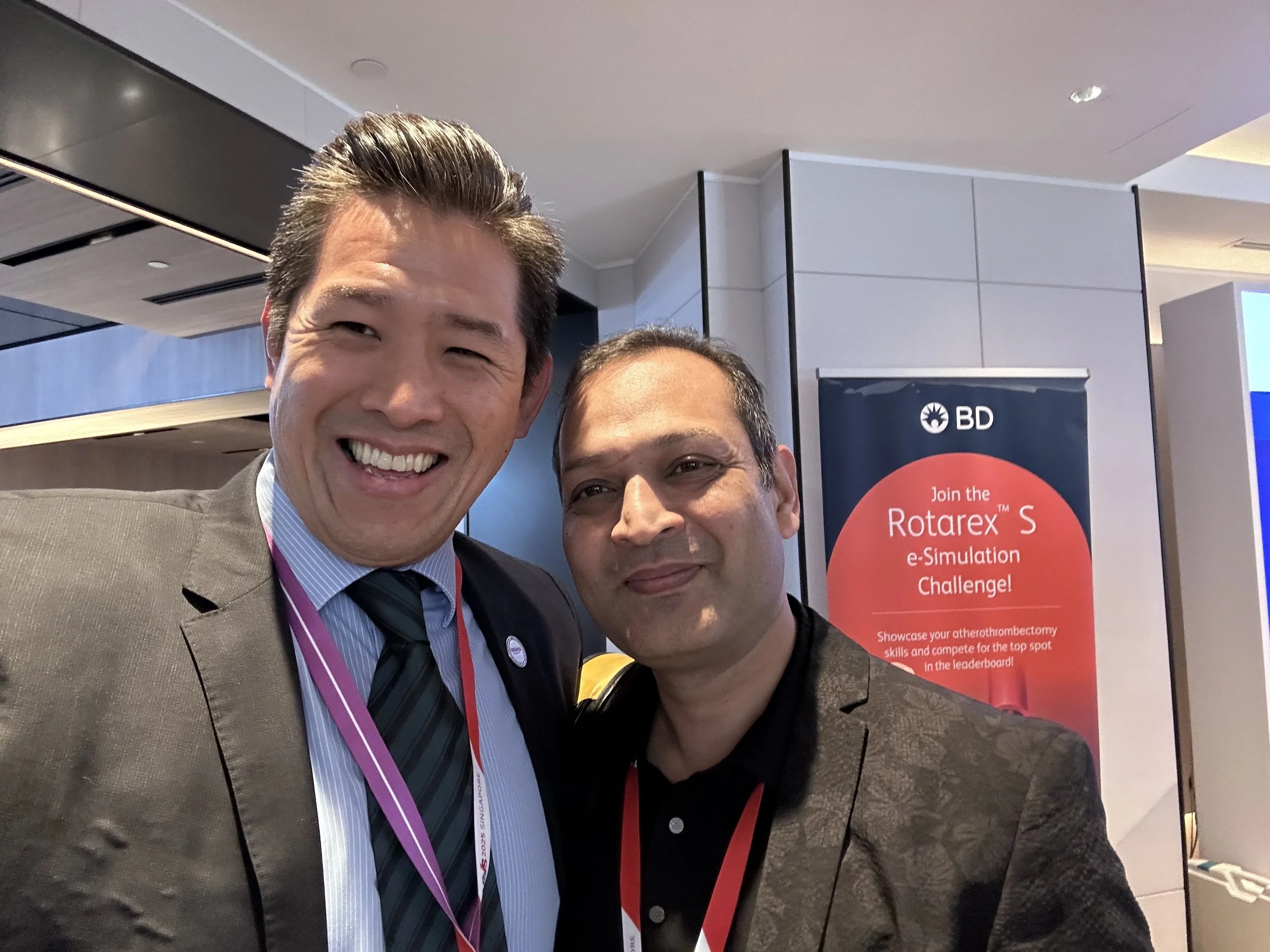
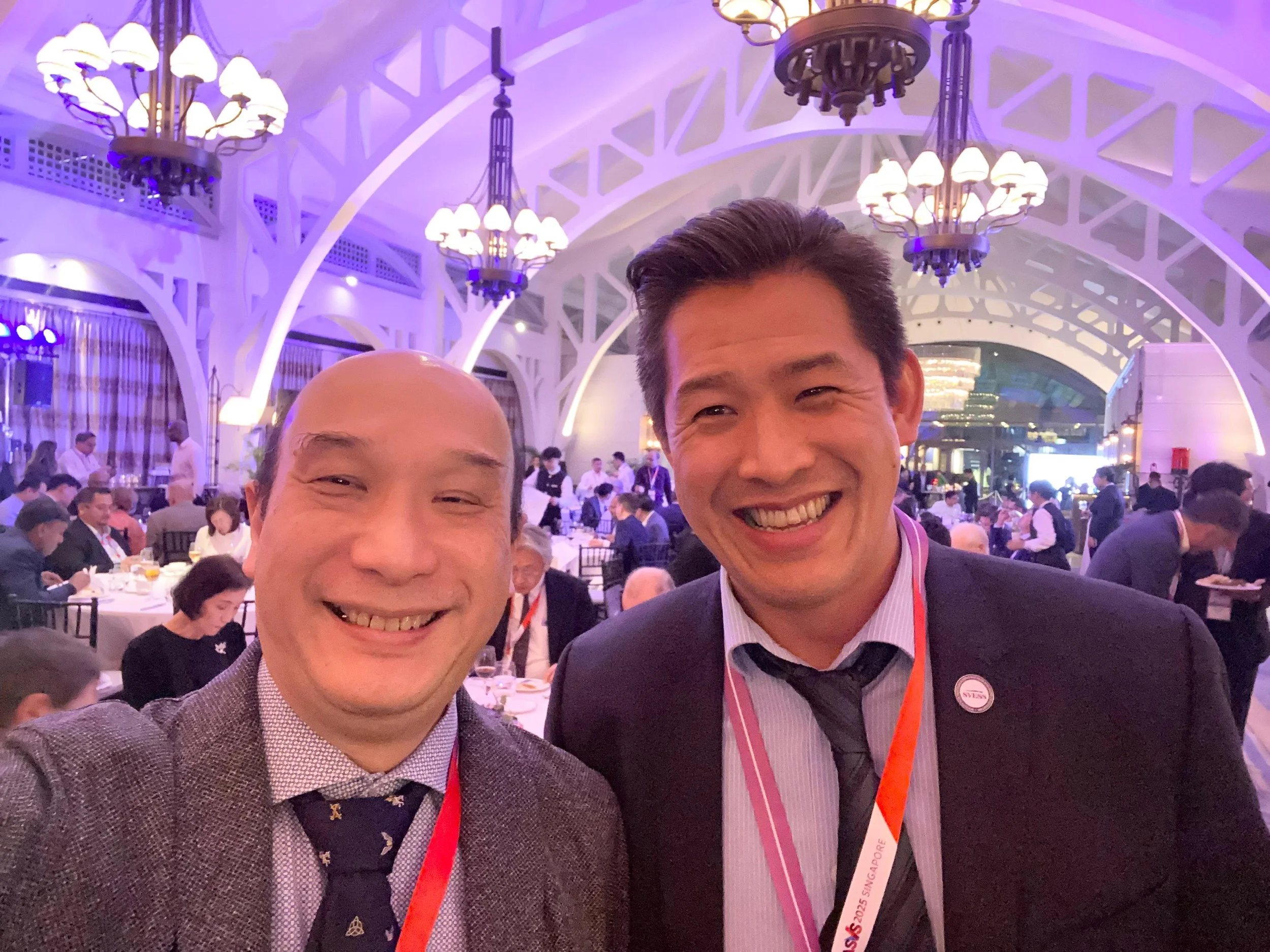
One of the pleasures of ASVS 2025 was reconnecting with friends and colleagues from across the world. Sharing experience gives us opportunities to question assumptions, discuss difficult decisions and learn from approaches used in different settings.
The photographs below capture some of those connections. They are a reminder that professional education also happens through conversation and collaboration.


What do these discussions mean for patients?
The message I took from these two talks is that established surgical skills and new technology both need thoughtful application. The aim is to understand the condition, explain the available options and agree a plan suited to the individual.
If you are discussing an aortic aneurysm with your surgeon, useful questions include:
What do my scans show, and how has the aneurysm changed?
Is surveillance appropriate, or is there a reason to consider repair now?
What are the benefits and risks of the options suitable for my anatomy?
What follow-up would I need after treatment?
I am grateful for the opportunity to share my work at ASVS 2025 and learn from colleagues. Continuing education remains part of how I approach vascular care, alongside careful assessment and clear communication with patients.

Sharing perspectives with fellow panellists at ASVS 2025 in Singapore.
Vascular assessment in Singapore
For questions about known aortic disease or treatment options, contact Andrew Choong Vascular Surgery at Mount Elizabeth Novena Specialist Centre, #09-22, 38 Irrawaddy Road, Singapore 329563. Previous scan reports and a medication list can help make the consultation more useful.
You can also explore my aortic awareness information and educational channels and updates.
This is a retrospective of a professional meeting and provides general education. Decisions about investigation or treatment require an individual clinical assessment.
Aortic Dissection Warning Signs: Think Aorta! Get a Contrast CT
Aortic dissection may cause sudden severe chest, back or abdominal pain, but it can also mimic other emergencies. Dr Andrew Choong explains warning signs, urgent assessment and the role of contrast CT when dissection is suspected.
Sudden severe chest, back or abdominal pain needs urgent emergency assessment. If you or someone with you has these symptoms, particularly with collapse, breathlessness or new weakness, do not wait at home or book a routine clinic visit. In Singapore, call 995 for emergency ambulance help.
In this video, ahead of Aortic Dissection Awareness Day, I explain why aortic dissection must remain a possibility when symptoms are sudden and serious. The message “Think Aorta” is a reminder to recognise the emergency and consider the diagnosis promptly.
What is an aortic dissection?
The aorta is the main artery carrying blood from the heart to the body. An aortic dissection occurs when a tear in its inner lining allows blood to enter and separate layers within the wall.
This can disrupt blood flow to important organs or limbs and can lead to other life-threatening complications. It is different from an aneurysm, which describes abnormal enlargement of an artery, although the two conditions can occur together. My aortic disease guide explains these differences in more detail.
What are the warning signs?
The classical symptom is pain that starts suddenly and is severe from the outset, often in the chest, back or abdomen. Some patients describe it as tearing or ripping; others describe the worst pain they have experienced.
However, you should not wait for a particular description of pain before seeking help. Aortic dissection does not always present in a textbook way, and a person may not use the words “tearing” or “ripping” at all.
Concerning features can include:
Sudden severe chest, back or abdominal pain.
Fainting, collapse or rapidly worsening illness.
New weakness, difficulty speaking or other stroke-like symptoms.
A sudden problem with circulation to a limb, such as pain, coldness or weakness.
These symptoms can also indicate other emergencies. The need for urgent care remains the same even when the exact cause is uncertain.
Why can aortic dissection be mistaken for something else?
In my video, I describe dissection as a clinical mimicker. Depending on the part of the aorta and its branches affected, it can resemble a heart attack, stroke, abdominal illness or limb circulation problem.
That is why the whole clinical picture matters. How suddenly did the symptoms start? Where is the pain? Are there changes in consciousness, movement or circulation? The emergency team brings these findings together rather than relying on a single symptom.
What does “get a contrast CT” mean?
When clinicians suspect an acute aortic problem, CT angiography is commonly used because it can rapidly show the aorta, the extent of a dissection and involvement of its branches. Contrast helps define the blood-filled channels and surrounding anatomy.
The phrase in my video is an awareness message about timely diagnosis. It is not advice for everyone with ordinary back pain to arrange a CT independently. The emergency team decides which imaging is appropriate, taking account of the clinical situation, stability, contrast history and other relevant factors. Alternative imaging may be needed in selected circumstances.
What should I tell the emergency team?
Describe when the symptoms began and whether they were sudden and severe. Mention known aortic disease, previous aortic procedures or a relevant family history if you know about them. Share your medication list and allergies when possible, without delaying the emergency call.
You do not need to prove that you have a dissection before asking for help. The priority is prompt assessment of a potentially serious event. Do not drive yourself if you are acutely unwell, and do not wait for a clinic message to be answered.
How is ongoing aortic care arranged in Singapore?
After an emergency has been assessed and treated, ongoing care may include blood-pressure management, specialist review and surveillance imaging. The plan depends on which part of the aorta is affected and the treatment already received.
For a non-emergency review of known aortic disease or a scan, contact Andrew Choong Vascular Surgery at Mount Elizabeth Novena Specialist Centre, #09-22, 38 Irrawaddy Road, Singapore 329563. You can also explore my aortic awareness information.
Awareness is useful when it leads to timely action: sudden, severe or otherwise concerning symptoms belong in an emergency assessment, not a period of observation at home.
World Patient Safety Day: Safer Vascular Care Starts Early
Safer vascular care starts with careful assessment and a clear treatment goal. Dr Andrew Choong explains why symptoms, scans, individual risks and patient preferences should guide decisions about monitoring, medication and surgery.
Safe vascular care begins before an operation or procedure is discussed. It starts with understanding the symptoms, examining the circulation, reviewing the right scans and asking what we are trying to prevent or treat.
For World Patient Safety Day, my message is that careful decisions matter at every stage. Aneurysms, blocked arteries, diabetic foot problems, deep vein thrombosis and carotid disease can have serious consequences, but their management needs to fit the individual patient.
Why does safety start with the assessment?
Similar symptoms can have different causes. Leg pain may relate to arterial circulation, veins, nerves or joints. Swelling may have several explanations. A scan abnormality also needs to be interpreted in context: we need to understand whether it explains the symptoms and whether it poses a risk that treatment can meaningfully reduce.
I begin by listening to what has changed and how it affects daily life. Examination, including pulse checks when appropriate, helps guide further investigation. Reviewing existing scans and previous treatment information can be as important as deciding whether another test is needed.
What problem is treatment meant to solve?
This is a practical question to ask in any vascular consultation. The goal might be to improve walking, support a wound's healing, reduce aneurysm-related risk or treat a blood clot. Different goals lead to different choices and different measures of success.
For example, assessment of peripheral arterial disease considers both walking symptoms and whether blood supply is sufficient for the foot. Assessment of aortic disease considers the part of the aorta affected, the scan findings, symptoms and overall health. A procedure is useful when its expected benefits justify its risks for that particular situation.
Can surveillance be an active treatment plan?
Yes. For some patients, monitoring and risk-factor management are the appropriate next steps. Surveillance means a planned review of the condition, with agreed follow-up and an explanation of what changes should trigger earlier reassessment. It does not mean ignoring a known problem.
Managing blood pressure, cholesterol, diabetes and smoking can be central to vascular care. The details depend on the condition and prescribed treatment. A good plan makes clear what needs monitoring, who is responsible for follow-up and what the patient should do if symptoms change.
Is the newest or least invasive procedure always safest?
No. Angioplasty, stents, endovascular repair, open surgery and clot-removal procedures each have roles. Their suitability depends on anatomy, disease severity, urgency, other medical conditions and the durability needed. A smaller incision alone cannot answer those questions.
Sometimes an endovascular approach is appropriate. Sometimes open surgery or a combination of techniques is a better fit. In other cases, an intervention would add risk without sufficient benefit. My aim is to explain the available options in relation to your condition rather than let a particular technology determine the plan.
How can patients take part in safer decisions?
Bring an up-to-date medication list, relevant scans and information about previous procedures. Tell the clinical team about allergies, kidney disease, previous reactions to contrast and concerns about recovery. These details help planning.
Questions worth asking include:
What is the diagnosis, and how certain are we?
What benefit would this treatment offer me?
What are the important risks and alternatives?
What happens if we monitor the condition first?
What follow-up and warning signs should I understand?
You should have an opportunity to discuss what matters to you, including mobility, independence and time away from work or family responsibilities. Clear explanations support a decision you understand.
When should assessment happen urgently?
Some changes cannot wait for scheduled review. Sudden severe chest or back pain, new stroke symptoms, collapse, serious breathlessness or a suddenly cold, painful or weak limb need emergency care. A deteriorating foot wound or new painful leg swelling also needs prompt assessment. If emergency ambulance help is needed in Singapore, call 995.
Where can I discuss my vascular care plan?
For a non-emergency assessment, scan review or second opinion, contact Andrew Choong Vascular Surgery at Mount Elizabeth Novena Specialist Centre, #09-22, 38 Irrawaddy Road, Singapore 329563. Careful assessment and a clear discussion of options are the starting point for choosing treatment at the right time.
Keyhole Vascular Treatment | Endovascular Surgery
Endovascular surgery treats blood vessels from the inside through a small access point. Dr Andrew Choong explains balloons, stents, stent-grafts and clot-removal devices, and why suitability depends on the condition and scan findings.
Endovascular treatment means treating a blood vessel from the inside. Through a small access point in an artery or vein, I can guide specialist instruments to the area that needs treatment. This is often described as “keyhole” or minimally invasive vascular surgery.
In this video, I introduce some of the tools used in modern vascular practice. The same small access point can support very different procedures, so it helps to understand the problem each tool is designed to address.
How do the instruments reach the blood vessel?
A small tube called a sheath provides access into the artery or vein. Guidewires and catheters can then be passed through it, with imaging used to guide their position. Depending on the condition, the treatment may involve balloons, stents, covered stent-grafts or devices designed to remove clot.
The access site and instruments are chosen for the anatomy and the intended procedure. The visible skin opening is only one part of the treatment: detailed planning and precise work take place inside the circulation.
How can a stent-graft treat an aneurysm?
An aneurysm is an abnormal enlargement of an artery. For a suitable aortic aneurysm, an endovascular stent-graft can create a channel for blood flow inside the diseased vessel. In my video, I describe this as a pipe within a pipe.
The graft is designed to seal against appropriate portions of the artery and exclude the aneurysm from the main blood flow. Whether this is possible depends on the shape, size and location of the aneurysm and the arteries used for access. You can read more in my aortic disease and aneurysm treatment guide.
Endovascular aneurysm repair also requires follow-up. Scans help check the graft and aneurysm over time, because treatment does not remove the need to monitor the remaining aorta.
What do balloons and stents do for narrowed arteries?
When an artery is narrowed or blocked, angioplasty uses a balloon to widen an appropriate segment. A stent may be used to support the treated vessel. The aim is to improve blood flow where the narrowing is causing a clinically important problem.
These techniques may be considered for selected patients with peripheral arterial disease, including some people with substantial walking limitation or threatened wound healing. Medication, risk-factor management and suitable exercise remain important; a scan showing narrowing does not automatically mean a stent is needed.
Can endovascular tools remove blood clots?
Some specialist devices can break up or remove clot through a catheter. Aspiration devices use suction; other mechanical systems work differently. The tool depends on what is blocking the vessel, where it is located and the patient's condition.
In selected cases of deep vein thrombosis or pulmonary embolism, a procedure to remove clot may be appropriate. It is not the standard answer for every blood clot. Anticoagulation and other treatments must be considered alongside the potential benefits and risks of an intervention.
Likewise, an artery blocked by longstanding plaque is not the same as a vessel containing a recent clot. Descriptions such as “drill” or “vacuum” are useful visual analogies, but they do not mean every blockage can or should be treated in the same way.
Does “keyhole” mean the procedure is minor?
No. A small access point can be used to treat a major vascular condition. Endovascular procedures can offer advantages for suitable patients, but they still carry risks and may require further treatment or ongoing surveillance. Some patients are better served by open surgery, a combined approach or medical management.
Before treatment, I consider the scans, overall health, clinical urgency and the outcome we are trying to achieve. I also discuss recovery, follow-up and alternatives so that the plan is understandable, rather than simply choosing the newest device.
Where can I discuss vascular treatment in Singapore?
For an individual opinion about vascular or endovascular treatment, contact Andrew Choong Vascular Surgery at Mount Elizabeth Novena Specialist Centre, #09-22, 38 Irrawaddy Road, Singapore 329563. Bring relevant scan reports and previous treatment information if available.
The tools are valuable when they solve the right problem. My role is to connect those options to your anatomy, symptoms and goals, and explain which approach is appropriate for you.
Poor Leg Circulation?
What happens during an assessment for poor leg circulation? Dr Andrew Choong explains the symptom history, foot examination and blood-flow tests that guide individual treatment for peripheral arterial disease in Singapore.
If you have been told that you may have poor circulation in your legs, the first step is to understand the blood-flow problem. The important questions are how much circulation is affected, where the disease is, and whether the foot or leg is at risk.
In this video, I explain what I look for during a vascular assessment for suspected peripheral arterial disease, or PAD. A treatment decision follows that assessment; it is not determined by the phrase “poor circulation” alone.
What is peripheral arterial disease?
Peripheral arterial disease occurs when arteries supplying the legs become narrowed or blocked, commonly because plaque has built up in their walls. Reduced blood flow may limit the oxygen available to the muscles during walking or, in more severe disease, affect the tissues even at rest.
PAD is an arterial problem. Other conditions, including venous disease, nerve problems and joint disorders, can also cause leg symptoms. Establishing the cause matters because their treatments are different.
Which symptoms can suggest poor arterial circulation?
A familiar pattern is discomfort or cramping in the calf, thigh or buttock that starts with walking and eases when you stop. This is called intermittent claudication. Some people also notice coldness, numbness or wounds that are slow to heal.
Symptoms that deserve particular attention include:
Walking pain that repeatedly limits the same everyday activities.
Foot or toe pain at rest, especially when persistent or occurring at night.
A wound or ulcer that is not healing.
Dark or black tissue, or a new change in foot colour or temperature.
These features need clinical assessment; none should be diagnosed from an online description alone. Diabetes can alter sensation, so a threatened foot may not always be as painful as expected.
What will I ask during the consultation?
The history helps connect the symptoms to what the leg is doing. How far can you walk? Where does the pain occur? Does it settle with rest, and does it return when you start again? Has the walking distance changed? Are there wounds, night pain or symptoms in the other leg?
I also ask about diabetes, kidney disease, smoking, heart disease and previous arterial treatment. Earlier angioplasty, stents or bypass operations can affect both the investigation and the options available. Bring your medication list and previous scan or operation information if you have it.
How are the legs and blood flow examined?
I examine the legs and feet, checking pulses, skin colour, temperature, wounds and signs of infection. The aim is to establish the likely circulation problem and identify whether the foot needs urgent attention.
Tests may include an ankle–brachial pressure measurement, which compares blood pressure at the ankle and arm, and duplex ultrasound to assess blood flow. Diabetes or kidney disease can make ankle-pressure results misleadingly high, so a normal result may not exclude PAD; toe-pressure testing or further assessment may be needed. Other tests or CT angiography may be appropriate when more detailed information is needed, particularly when planning a procedure. The choice depends on the clinical question rather than ordering every available scan.
Does PAD always need angioplasty or surgery?
No. Many patients need a plan built around prescribed medication, management of cardiovascular risk factors and an appropriate structured exercise programme. Stopping smoking and controlling diabetes, blood pressure and cholesterol are important parts of care.
For selected patients, angioplasty, stenting or bypass surgery may improve circulation. A procedure may be considered for symptoms that remain substantially limiting despite suitable treatment, or when blood supply threatens wound healing or limb survival. I weigh the artery pattern, wound risk, expected benefit and overall health together.
The goal is more than opening a narrowed artery on a scan. It is to improve function, support healing when needed and reduce the risk of complications.
When is poor circulation an emergency?
A suddenly painful, cold, pale, numb or weak limb needs immediate emergency assessment. Seek urgent care rather than waiting for a routine appointment. A non-healing wound, black toe or persistent rest pain also needs prompt assessment; do not try to exercise through a threatened or infected foot.
Where can I arrange a circulation assessment in Singapore?
For non-emergency symptoms or review of known PAD, contact Andrew Choong Vascular Surgery at Mount Elizabeth Novena Specialist Centre, #09-22, 38 Irrawaddy Road, Singapore 329563. I will assess your symptoms, examination and blood-flow findings together to discuss a treatment plan suited to you.
Varicose veins: scan first, treatment second
Similar-looking varicose veins can have different causes beneath the surface. Dr Andrew Choong explains how vascular ultrasound identifies venous reflux and guides a treatment plan that matches your symptoms and scan findings.
The most suitable treatment for varicose veins cannot be chosen simply by looking at the veins on the surface. A proper assessment, including vascular ultrasound when planning treatment, helps identify the underlying problem and guides what to do next.
In this video, I explain why I start with the question of where abnormal blood flow is coming from. Two people may have very similar-looking veins but need different treatment plans.
Why are visible varicose veins only part of the picture?
Veins return blood towards the heart. In the legs, one-way valves help prevent blood from falling backwards between contractions of the leg muscles. When those valves do not work properly, blood can flow in the wrong direction. This is called venous reflux.
The bulging veins you can see may be the result of reflux elsewhere in the venous system. Treating a visible vein without understanding that pattern may leave an important source of the problem untreated. My guide to varicose veins and venous disease explains how reflux can contribute to aching, heaviness, swelling and skin changes.
What does a vascular ultrasound scan show?
A duplex ultrasound examines both the structure of the veins and the way blood moves through them. It helps map which veins are involved and where reflux occurs. The findings are interpreted alongside symptoms, examination and previous treatment rather than in isolation.
This mapping can distinguish problems in the main superficial veins, their branches and the deeper venous system. These distinctions matter: a visible cluster of veins does not automatically tell us which vessel should be treated or whether it is the main cause of the symptoms.
The scan therefore provides information that a photograph cannot. It gives the treatment discussion a basis beyond appearance alone.
Why might two similar-looking legs need different treatments?
One person may have reflux in a superficial vein that is suitable for an endovenous procedure. Another may have a different reflux pattern, previous vein treatment or additional venous problems that change the plan. Sometimes the visible veins are not the main issue.
I also consider what is troubling the patient. Discomfort during work, swelling at the end of the day, troublesome itching and skin changes raise different questions from appearance alone. Understanding the effect on daily life helps us decide what improvement treatment should aim to achieve.
Which treatments might follow the scan?
Depending on the findings, options can include endovenous laser treatment, radiofrequency ablation, medical glue or ultrasound-guided foam sclerotherapy. These approaches work in different ways to close an appropriate abnormal superficial vein so that blood can return through other functioning veins.
Some patients need a different procedure or a combination of treatments. Others may benefit from conservative management and review. No single method is right for every leg, and finding reflux does not mean everyone must immediately undergo an intervention.
The discussion should cover the expected benefit, possible complications, alternatives and follow-up. It should also explain whether treating the main source of reflux and treating visible branches would happen together or in stages.
Further guides cover microphlebectomy for surface branches and ligation and stripping, including selection, risks and recovery.
What should I ask before agreeing to varicose vein treatment?
Useful questions include:
Where is the reflux coming from, and does it explain my symptoms?
Which vein would be treated, and why?
What alternatives are suitable for my scan findings?
What recovery, follow-up and possibility of further treatment should I expect?
A clear plan connects the ultrasound findings to your symptoms and goals. The name of a device or a treatment alone cannot provide that explanation.
Read our vascular duplex ultrasound guide for more about the scan, preparation and how results are discussed.
When should I arrange an assessment in Singapore?
Consider vascular review if varicose veins cause persistent aching, heaviness, swelling, itching or skin changes, or if you are considering treatment. An ulcer, bleeding vein or sudden painful swelling needs prompt medical attention; sudden swelling should not simply be assumed to be ordinary varicose veins.
For an individual assessment, contact Andrew Choong Vascular Surgery at Mount Elizabeth Novena Specialist Centre, #09-22, 38 Irrawaddy Road, Singapore 329563. I will relate your symptoms and scan findings to the options available. Scan first, treatment second: the aim is a plan that fits the actual problem.
What Is Linked to 11 Million Deaths a Year? Sepsis, Foot Wounds and Poor Circulation
A foot wound can become dangerous when infection and poor circulation occur together. Dr Andrew Choong explains sepsis warning signs, why diabetes can mask pain, and when a worsening wound needs urgent assessment in Singapore.
Sepsis happens when the body's response to an infection begins to damage its own tissues and organs. It can become life-threatening quickly. In this World Sepsis Day video, I explain why a worsening foot wound deserves particular attention when someone has diabetes or poor circulation.
As a vascular surgeon in Singapore, I look at two linked questions: is the wound infected, and is enough blood reaching the foot for it to heal? Both matter when deciding how urgently someone needs care.
What does the figure of 11 million deaths mean?
The figure in my video reflects a major global estimate: approximately 48.9 million sepsis cases and 11 million sepsis-related deaths worldwide in 2017, reported in research published in 2020. These figures describe the scale of the problem; they are not a new count for 2026 or a prediction of what will happen to an individual patient.
Sepsis can arise from many infections. A foot wound is one possible source, particularly when infection spreads or a person's ability to fight infection and heal is impaired.
Why can a small foot wound become serious?
A small break in the skin can allow infection to develop. Diabetes, kidney disease and poor arterial circulation can complicate the situation. A history of smoking also matters because it increases the risk of vascular disease. The appearance of the wound alone does not tell the whole story.
When peripheral arterial disease reduces blood flow, the tissues may not receive enough oxygen and nutrients to support healing. Antibiotics and appropriate dressings may be necessary, but they do not automatically correct a circulation problem. Infection control and assessment of blood supply need to be considered together.
Can a diabetic foot infection be serious without much pain?
Yes. Diabetes can damage the nerves that normally warn us about an injury. A wound or infection may therefore hurt less than expected. Little pain is not reliable reassurance, especially if the foot looks different, the wound is worsening or the person feels unwell.
This is why I encourage patients and families to notice changes rather than wait for severe pain. A new wound, spreading redness, increasing swelling, discharge or a change in skin colour needs attention, even when walking remains possible.
Which warning signs need urgent help?
A foot wound needs prompt medical assessment if it develops pus, worsening redness or swelling, severe pain, black tissue or a rapid change in appearance. In someone with diabetes or known poor circulation, do not wait for a wound to become large before seeking help.
Possible sepsis is an emergency. Warning signs may include:
New confusion, unusual drowsiness or sudden deterioration.
Severe shivering, fever, or feeling markedly unwell.
Breathlessness or unusually rapid breathing.
Passing very little urine.
Mottled, pale, blue or otherwise discoloured skin.
Not everyone has every sign. If someone with an infection becomes seriously unwell, seek emergency care immediately; in Singapore, call 995 when emergency ambulance help is needed. Do not wait for a routine clinic appointment.
How does a vascular assessment help?
I assess the wound alongside the person's symptoms, medical history, foot temperature, skin changes and pulses. When indicated, blood-flow tests or vascular ultrasound help establish whether narrowed or blocked arteries are contributing to poor healing. Further imaging depends on the findings and the treatment being considered.
The plan may involve infection treatment, wound care, pressure relief and management of diabetes and other risk factors. Some patients also need a procedure to improve circulation. The right sequence depends on the severity of infection, the blood supply and the person's overall health.
For more on ongoing care, read our diabetic foot and wound care guide. Suspected sepsis still needs immediate emergency assessment, as explained above.
Where can I discuss a foot wound or circulation concern in Singapore?
For a non-emergency vascular assessment, contact Andrew Choong Vascular Surgery at Mount Elizabeth Novena Specialist Centre, #09-22, 38 Irrawaddy Road, Singapore 329563. I can assess whether poor circulation is affecting healing and discuss an appropriate treatment plan.
This article accompanies my video for general patient education. A worsening wound needs an individual assessment, and suspected sepsis requires immediate emergency care.
Can Walking Improve Your Circulation???
Walking can help arteries and veins in different ways. Dr Andrew Choong explains structured walking for stable claudication, the calf muscle pump, practical ways to build a routine and warning signs that should never simply be walked off.
Walking can support both arterial and venous circulation, but it helps them in different ways. For people with assessed, stable claudication, structured walking can improve walking ability. For venous symptoms, each step activates the calf muscles and helps move blood back towards the heart.
In this video, I explain these two benefits and the warning signs that should never simply be “walked off”. The right exercise plan depends on the cause of your symptoms, your overall health and whether it is safe for you to walk.
How can walking help arterial disease?
Claudication is cramping, aching or tightness in the calf, thigh or buttock that develops with activity and improves with rest. When it is caused by peripheral arterial disease (PAD), narrowed arteries cannot meet the muscles’ increased demand for oxygen during walking.
Regular training can improve fitness and how effectively the leg muscles use oxygen, helping some people walk further before symptoms limit them. This does not mean that a blocked artery has simply opened or that everyone will improve at the same rate.
Exercise is one part of PAD care. Smoking cessation, management of cholesterol, blood pressure and diabetes, and prescribed medicines also matter because PAD is linked to wider cardiovascular risk.
What is the walk–rest–repeat approach?
For suitable people with stable claudication, a structured programme often uses intervals of walking and rest. In the video, I describe this pattern:
Walk until the familiar discomfort becomes moderate. Use the level of effort agreed with your clinician.
Stop and rest until it settles. Allow the discomfort to ease before starting again.
Resume walking and repeat. Build consistency within an individual programme rather than force a particular distance.
A supervised or clinician-supported programme can help set a suitable pace and monitor progress. This approach is for an assessed condition; unexplained new pain should first be investigated. Severe pain, a sudden change or symptoms at rest should not be treated as a training target.
How does walking help the veins?
Leg veins return blood towards the heart. When your calf muscles contract, they squeeze the veins and help propel blood upwards. One-way valves support this flow and limit backward movement.
Walking therefore activates the calf muscle pump, unlike remaining still for long periods. It can help some people with venous heaviness, aching or swelling. If there is venous reflux or varicose veins, movement may be part of symptom management, but it does not repair faulty valves.
Compression may also be appropriate for selected patients. The type and pressure should be advised according to the venous problem, arterial circulation and other health conditions.
How can you make walking a sustainable habit?
Choose a route and pace that suit your mobility and balance. Comfortable, well-fitting footwear matters, particularly if you have diabetes or reduced sensation. Check your feet for rubbing, blisters or wounds rather than relying only on pain to alert you.
In Singapore’s heat, a cooler time of day or a sheltered route may make activity more comfortable. Build the routine around your circumstances and any medical advice, including fluid restrictions if you have them.
A simple note of your walking time, symptoms and recovery can help you and your clinician see whether the pattern is improving or changing. Walking can also support glucose control, heart health and general conditioning, although it does not replace prescribed treatment.
When should you stop and seek medical help?
Seek urgent assessment for new pain at rest, a non-healing wound, blackened toes or one-sided painful swelling. Swelling with warmth or tenderness may need investigation for DVT, not a longer walk.
Sudden severe leg pain with coldness, numbness or weakness requires emergency assessment. Chest pain, sudden breathlessness or collapse also need emergency care. In Singapore, call 995 for a life-threatening emergency rather than waiting for a routine appointment.
Assessment and Treatment Guides
For more on the next steps, see vascular duplex ultrasound assessment and leg angioplasty and stenting for selected patients. The appropriate next step depends on clinical assessment.
Planning safe walking with a vascular condition
If leg symptoms are limiting your daily activity, contact Andrew Choong Vascular Surgery to discuss assessment at Mount Elizabeth Novena Specialist Centre, #09-22, 38 Irrawaddy Road, Singapore 329563. Understanding the cause helps us decide how walking fits into your care.
This article provides general education and does not replace an individual medical assessment.
DVT Warning: Red, hot, swollen painful leg
New one-sided leg swelling, pain or warmth may be a DVT and needs urgent assessment. Dr Andrew Choong explains possible symptoms, pulmonary embolism warning signs, risk factors and how venous ultrasound helps investigate a suspected blood clot.
A new swollen, painful or unusually warm leg, especially on one side, needs urgent medical assessment. It may be a deep vein thrombosis, or DVT: a blood clot in a vein deep inside the leg. Not everyone has every symptom, and you should not wait for a complete checklist before seeking help.
In this video, I explain why this combination matters, which situations can increase the risk of a clot and how an ultrasound scan helps investigate it. The message is straightforward: do not dismiss a sudden change as ordinary tiredness or your usual leg swelling.
What symptoms can a DVT cause?
Possible symptoms include:
Swelling: commonly affecting one leg more than the other.
Pain or tenderness: often in the calf or thigh, without an obvious explanation.
Warmth: an affected area that feels hotter than usual.
A change in skin colour: including redness or darker colouring, which may be less obvious on some skin tones.
Other conditions, such as infection, injury or inflammation, can produce similar symptoms. Some people with DVT have few or no obvious signs. This is why appearance alone cannot confirm or exclude a clot. Our DVT information page explains the condition in more detail.
Why can a clot in the leg affect the lungs?
A DVT can interfere with blood drainage from the leg. If part of the clot breaks away, it can travel through the circulation and lodge in the blood vessels of the lungs. This is called a pulmonary embolism (PE), which can be life-threatening.
Sudden breathlessness, chest pain, coughing blood, fainting or collapse require emergency assessment. These symptoms can occur even without obvious leg symptoms. In Singapore, call 995 for a life-threatening emergency. Do not wait for a clinic appointment or an online reply.
Who has a higher risk of DVT?
In the video, I highlight cancer, some blood conditions, long-haul travel, periods of immobility and major orthopaedic surgery. These situations may increase the tendency for blood to clot or reduce its movement through the leg veins.
Previous DVT, pregnancy and the period after childbirth, and some hormone treatments are also relevant. Several risks can occur together. However, DVT can happen without an obvious trigger, so the absence of a recent flight or operation is not a reason to ignore symptoms.
Long journeys by car, bus or train can also involve prolonged sitting. The concern is not unique to flying; limited movement and a person’s underlying risk both matter.
How is a suspected DVT assessed?
A clinician will ask about the symptoms, examine the leg and review your risk factors. A venous duplex ultrasound is a common first-line imaging test. It examines the deep veins and helps identify a clot and its location.
Depending on the clinical assessment, blood tests or further imaging may also be needed. If symptoms persist or change after an initial assessment, follow the advice given about reassessment. A scan result needs to be interpreted alongside the full clinical picture.
What happens if a DVT is confirmed?
Treatment commonly involves anticoagulant medicines, often called blood thinners. They help stop the clot extending and reduce the risk of further clots while the body deals with the existing clot. The medicine and duration depend on factors such as the clot’s location, cause and bleeding risk.
Some patients need additional specialist treatment. Follow-up also considers ongoing swelling or discomfort and whether there are underlying factors to address. A new suspected clot needs urgent care, rather than treatment based only on information in a video.
What can you do before a long journey?
Move your legs regularly and take opportunities to walk when safe. Keeping normally hydrated is sensible, but drinking water alone should not be regarded as protection against a travel-related clot.
If you have had a clot, recent surgery, cancer or another significant risk factor, discuss travel precautions with your clinician beforehand. Compression stockings or preventive medicines are appropriate for selected people and should be individually advised.
Related DVT Guides
Read more about DVT treatment in Singapore, pulmonary embolism and vascular duplex ultrasound. These guides support the urgent-care advice above; do not delay assessment for new symptoms.
DVT assessment in Singapore
Seek urgent medical care for new one-sided painful swelling. For non-emergency questions about previous DVT or ongoing venous care, contact Andrew Choong Vascular Surgery at Mount Elizabeth Novena Specialist Centre, #09-22, 38 Irrawaddy Road, Singapore 329563.
This article provides general education and does not replace an individual medical assessment.